Welcome to Companion Animal Hospital
Companion Animal Hospital of Traverse City is a veterinary practice for dogs, cats, rabbits, and pocket pets with high standards, practicing extraordinary medicine with over 40 years of experience.

Shop Now
Our online store is your go-to destination for all your pet’s health and wellness needs.
Request Appointment
Click below to schedule an appointment at Companion Animal Hospital in Traverse City!
New Client
Welcome to our client family! Click below for a snapshot of what to expect from your first visit.
Companion Animal Hospital's Vision
Companion Animal Hospital was founded by Dr. Marcia Izo in April 2002. Dr. Izo and her husband, Terry, built the state-of-the-art facility in order to provide an exceptional experience for both patients and their owners. Today, our veterinarians strive to find the best solutions for your pet(s), while developing long-lasting relationships with our clients and facilitating a trusting and familiar atmosphere for all companions. Dr. Izo’s vision remains the guidepost for everything we do and how we do it!


Complete Veterinary Services in Traverse City, MI
We are a full-service veterinary hospital specializing in the compassionate care of your furry family members throughout their lives. Our clients and patients are treated very well at our hospital and always with respect!
Wellness & Preventative Care
The most important part of preventive care is the regular, very thorough, nose-to-tail Comprehensive Physical Examination.

In-House Diagnostics
As dogs and cats age, they tend to develop more health issues similar to humans, but at a faster rate. Our goal is to identify those problems early.
Surgery
We provide extensive surgical procedures, ensuring your furry family receives the best care with our skilled soft tissue surgeons, advanced facilities.

Pocket Pet Care
We know the importance of our household rabbits and small pocket pet friends. Our veterinarians treat rabbits, guinea pigs, hedgehogs, chinchillas, hamsters, gerbils, mice and rats.

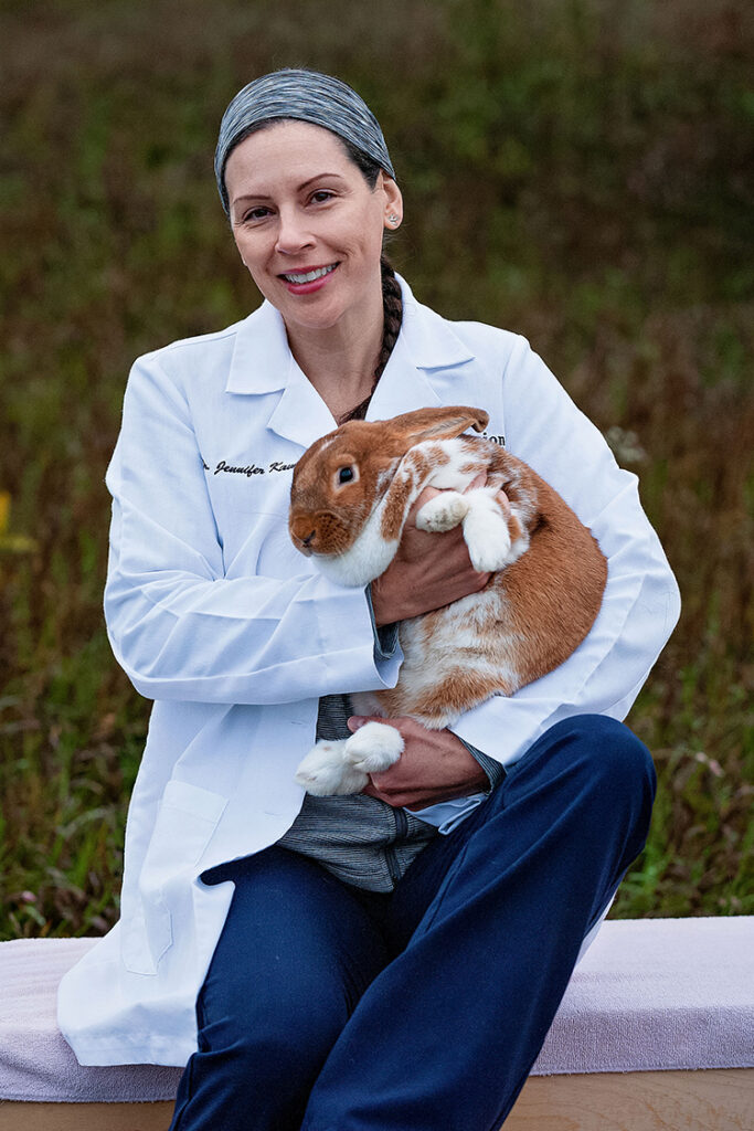
Meet Our Veterinary Team
At Companion Animal Hospital in Traverse City, MI, we take pride in being your trusted partners in pet care. Our dedicated veterinary team is committed to providing compassionate, comprehensive, and cutting-edge medical services for a wide variety of furry and feathered companions. Led by two accomplished veterinarians, Dr. Jennifer E. Kavran and Dr. Rebecca DeSimone, our team is driven by a shared passion for animal health and well-being.
Thank You for Your Kind Words
